Description
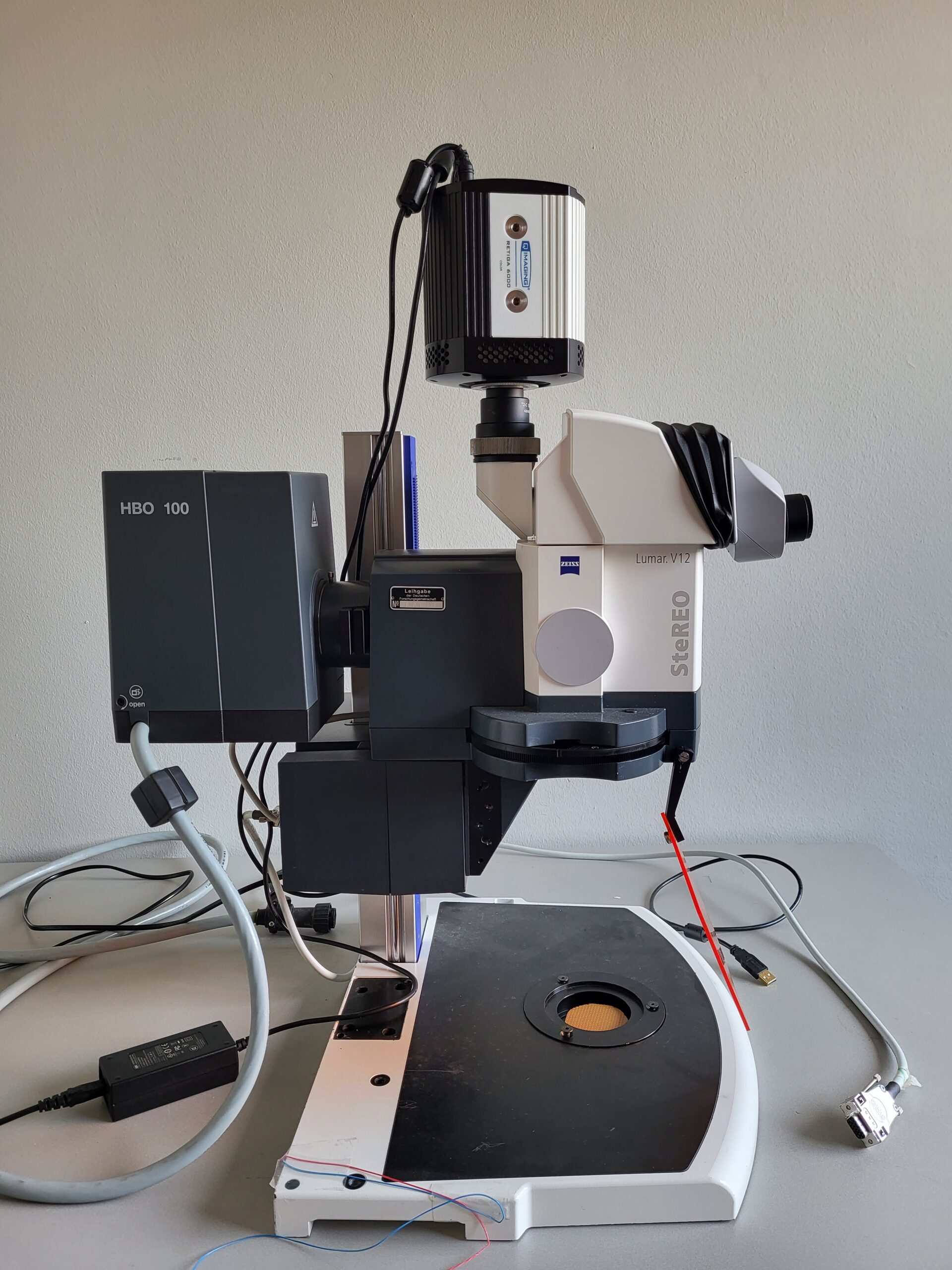
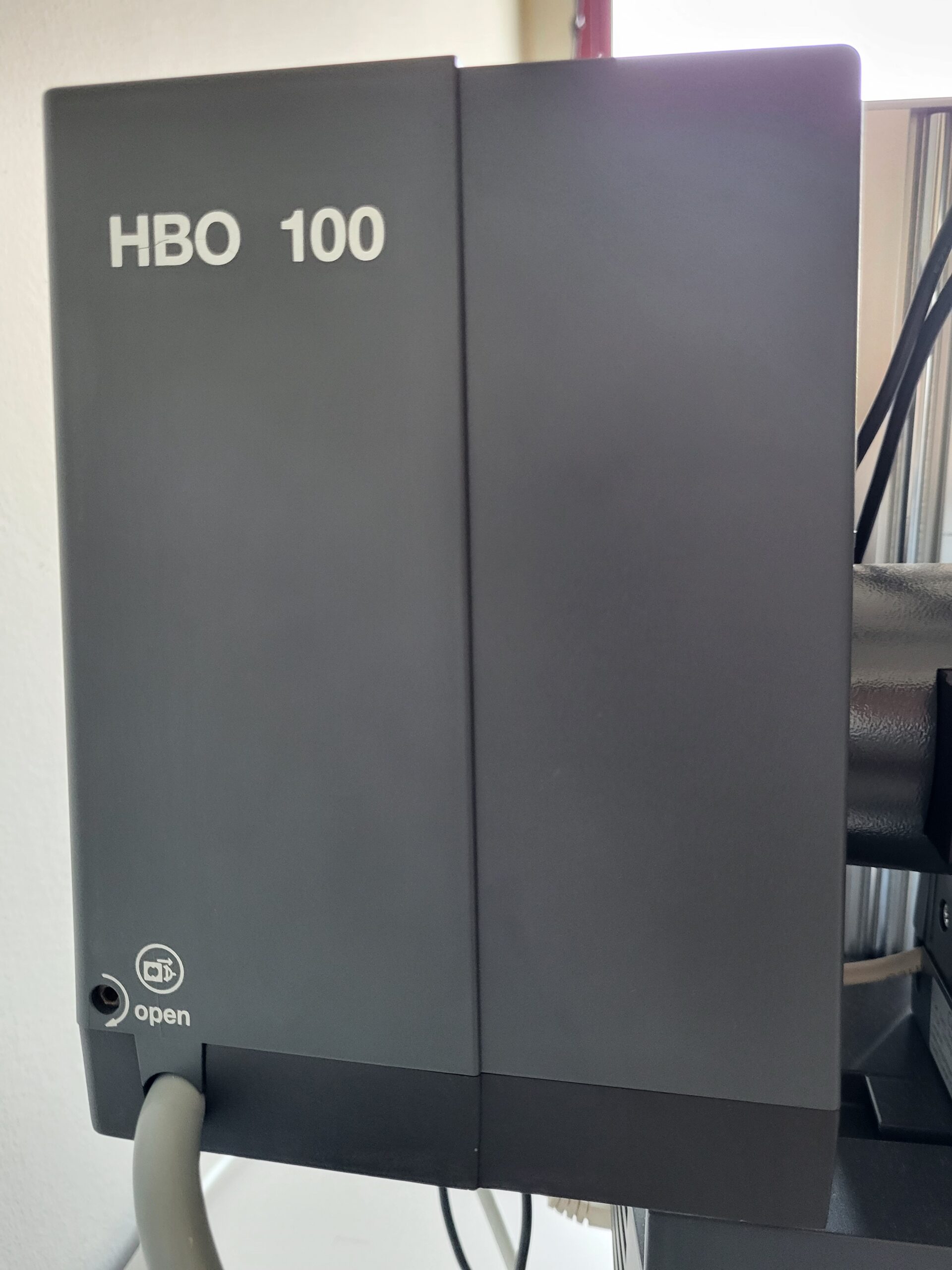
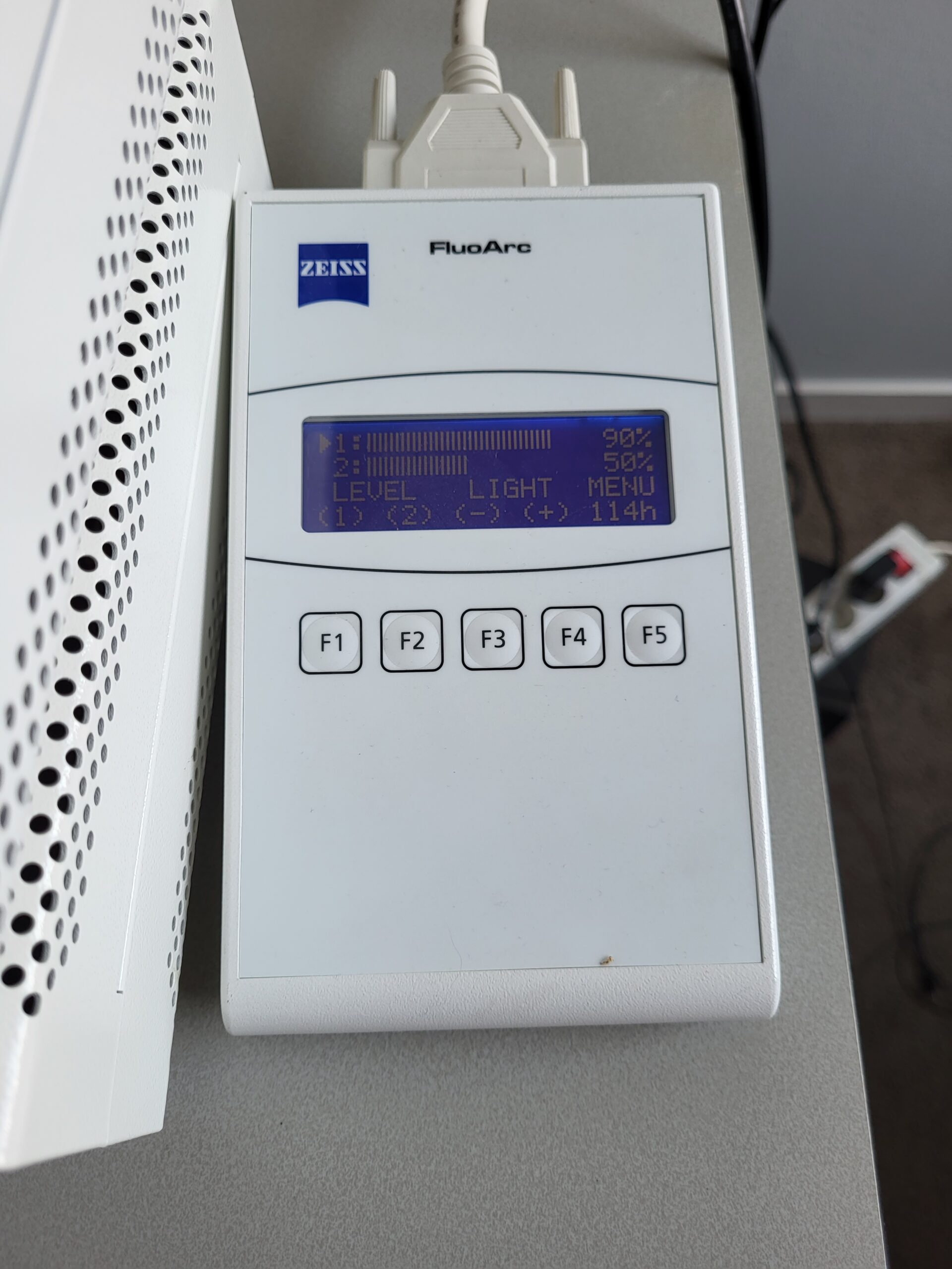
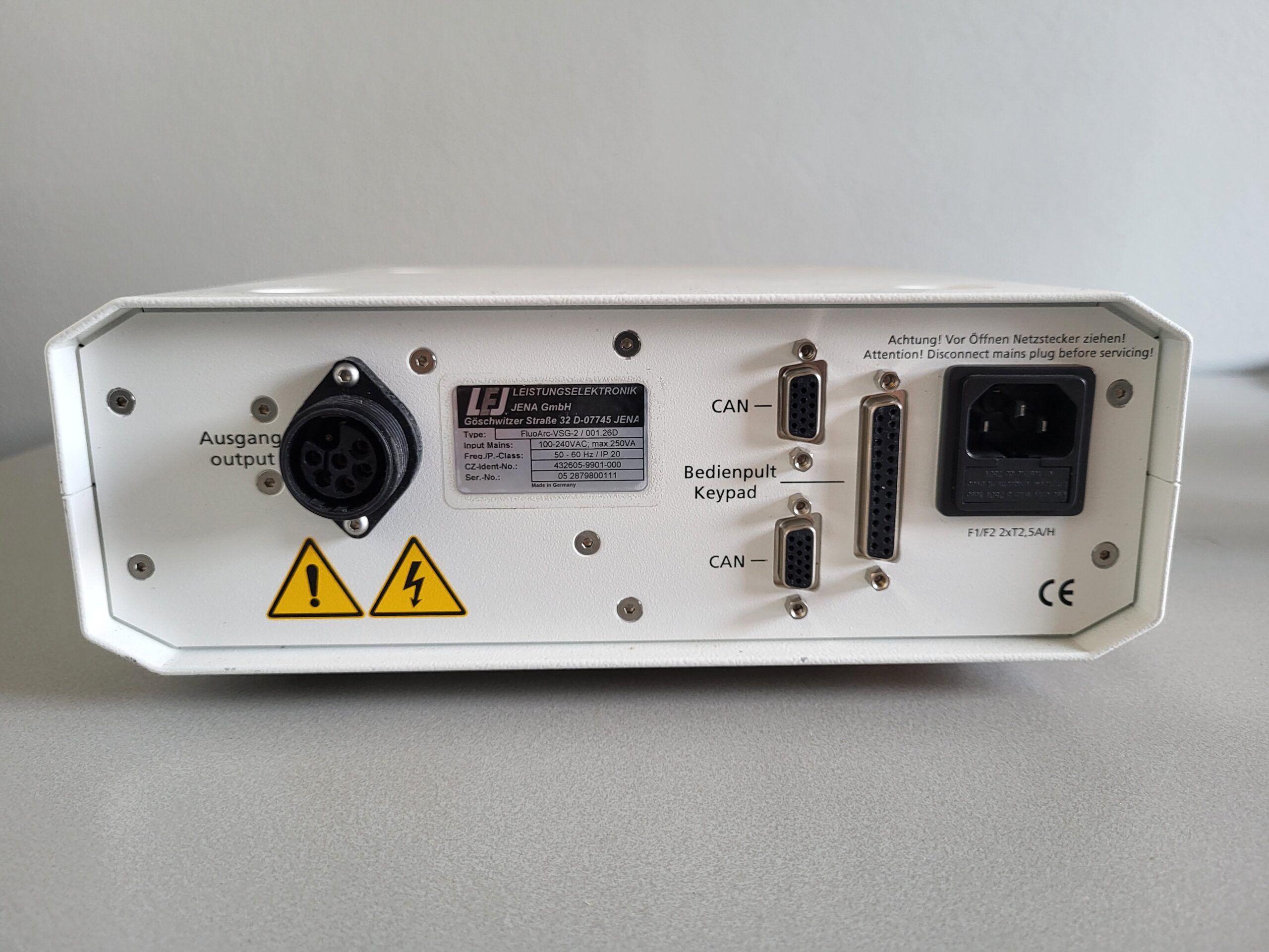
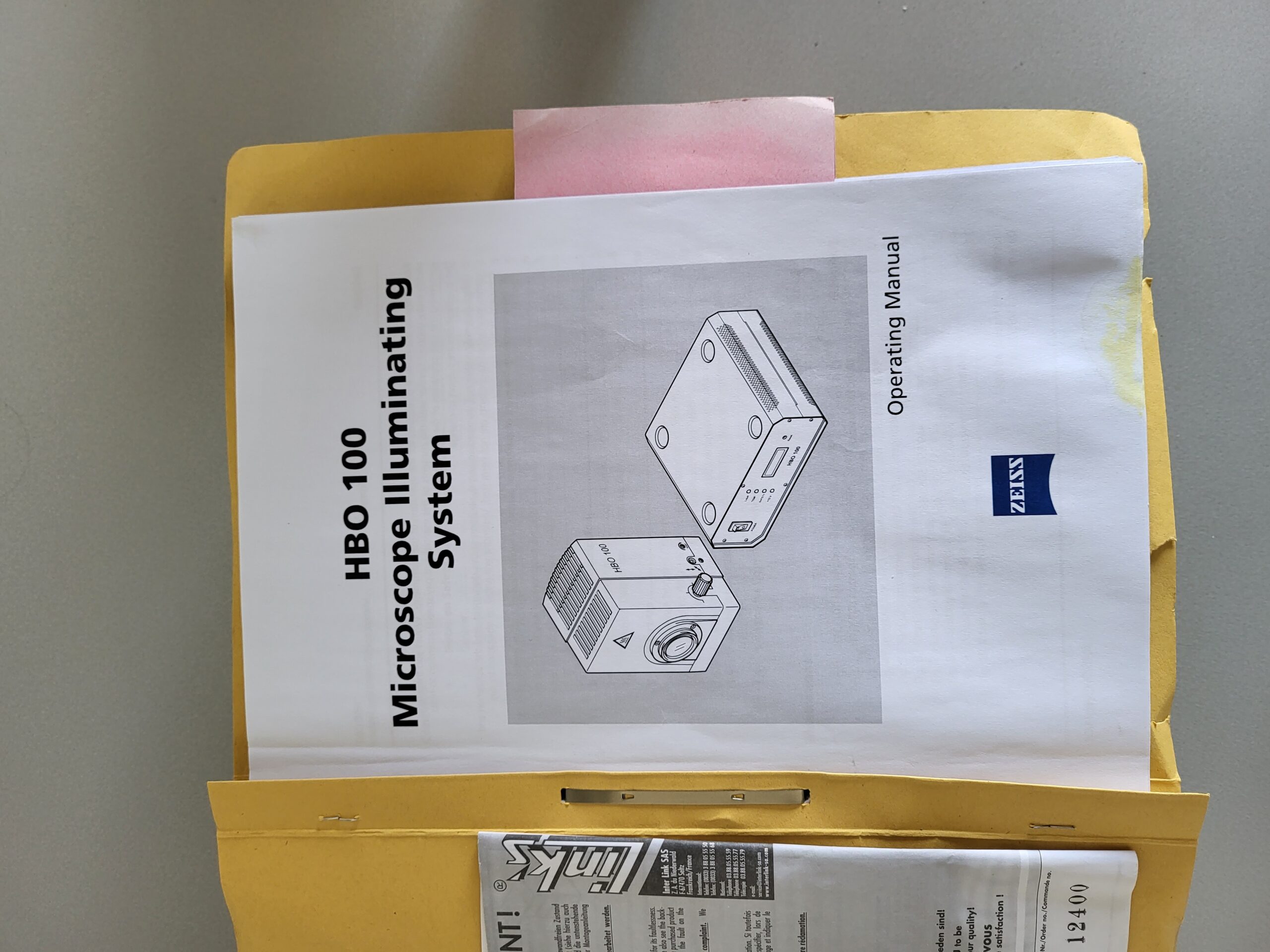
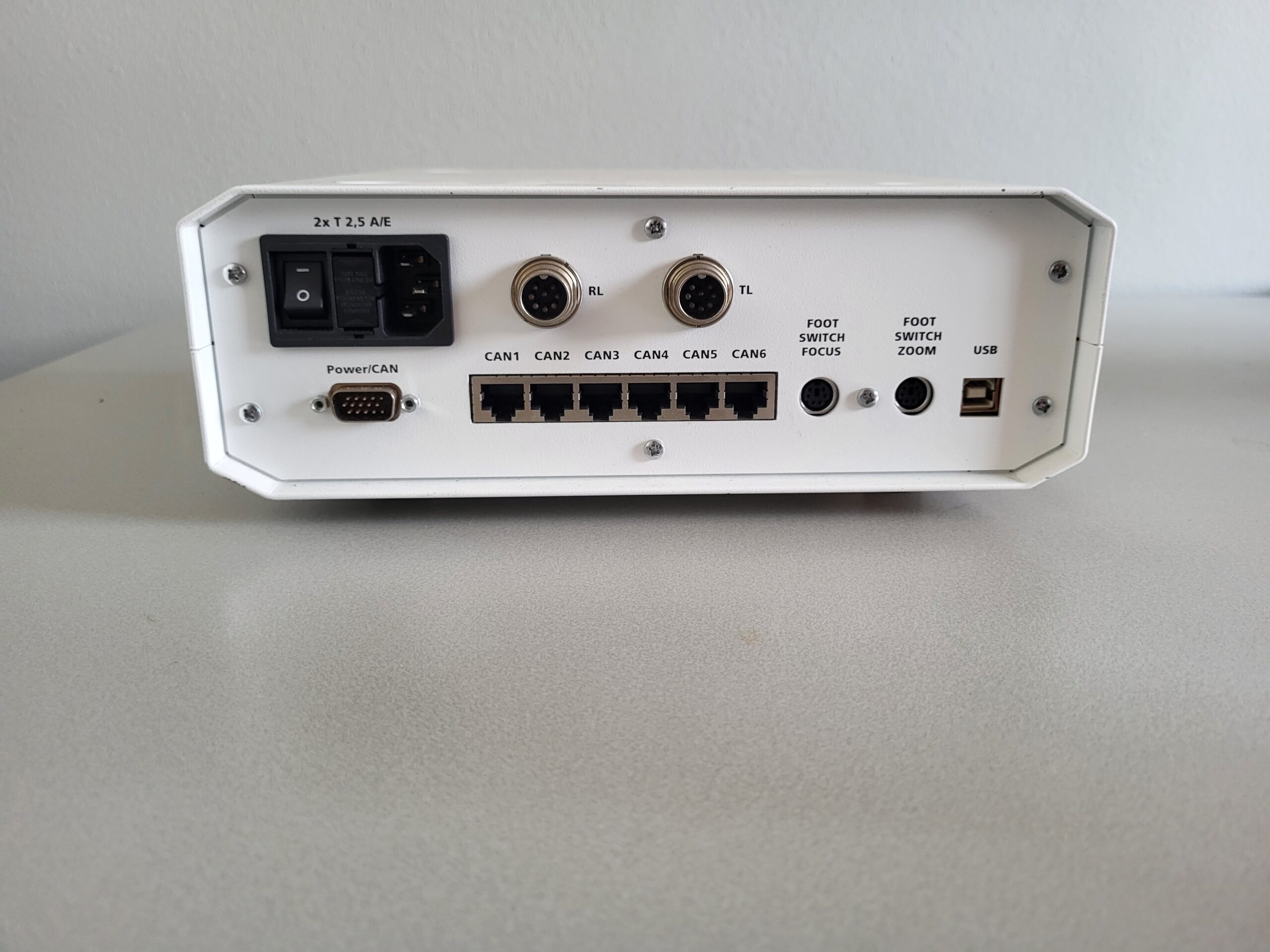
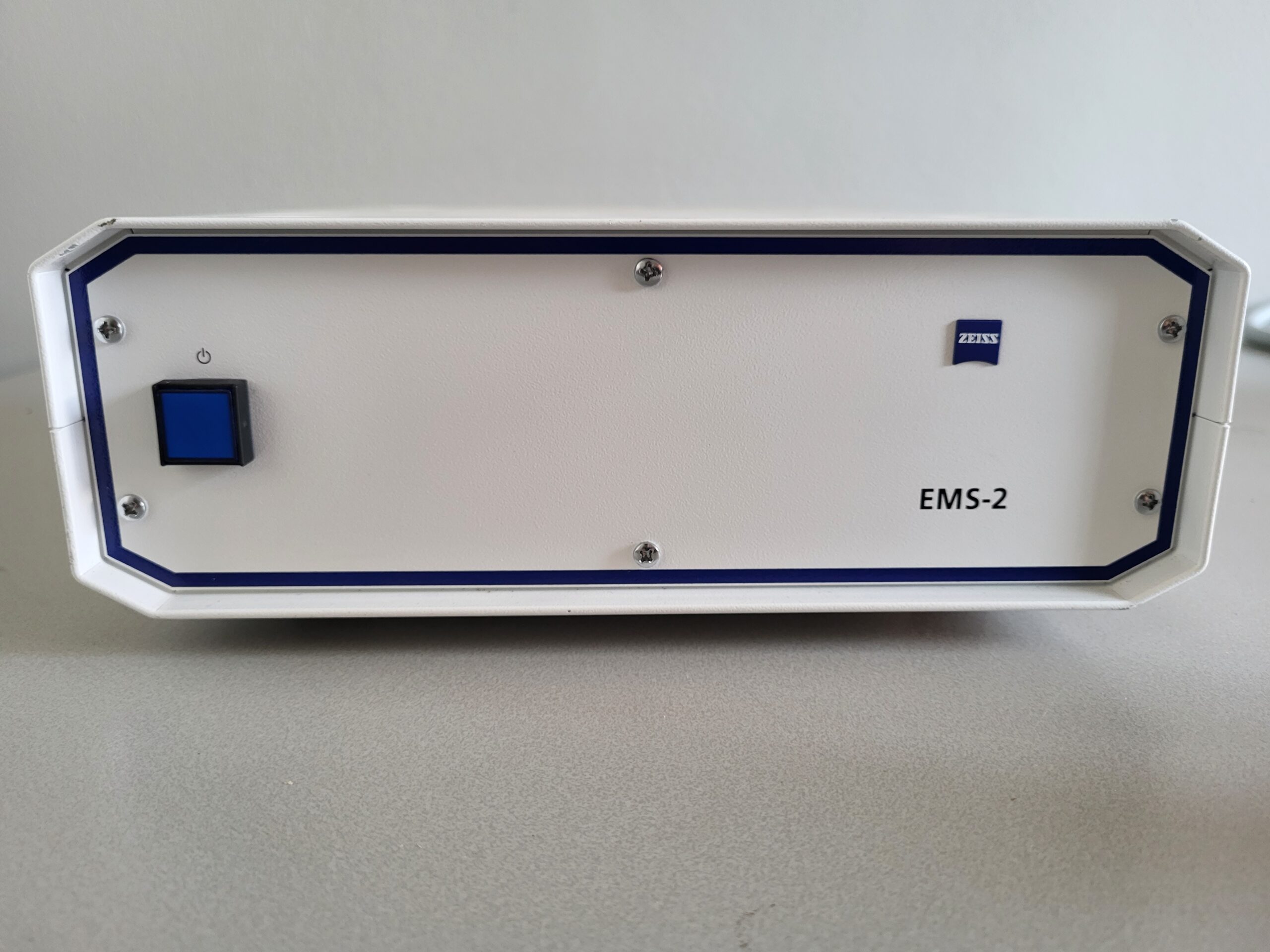
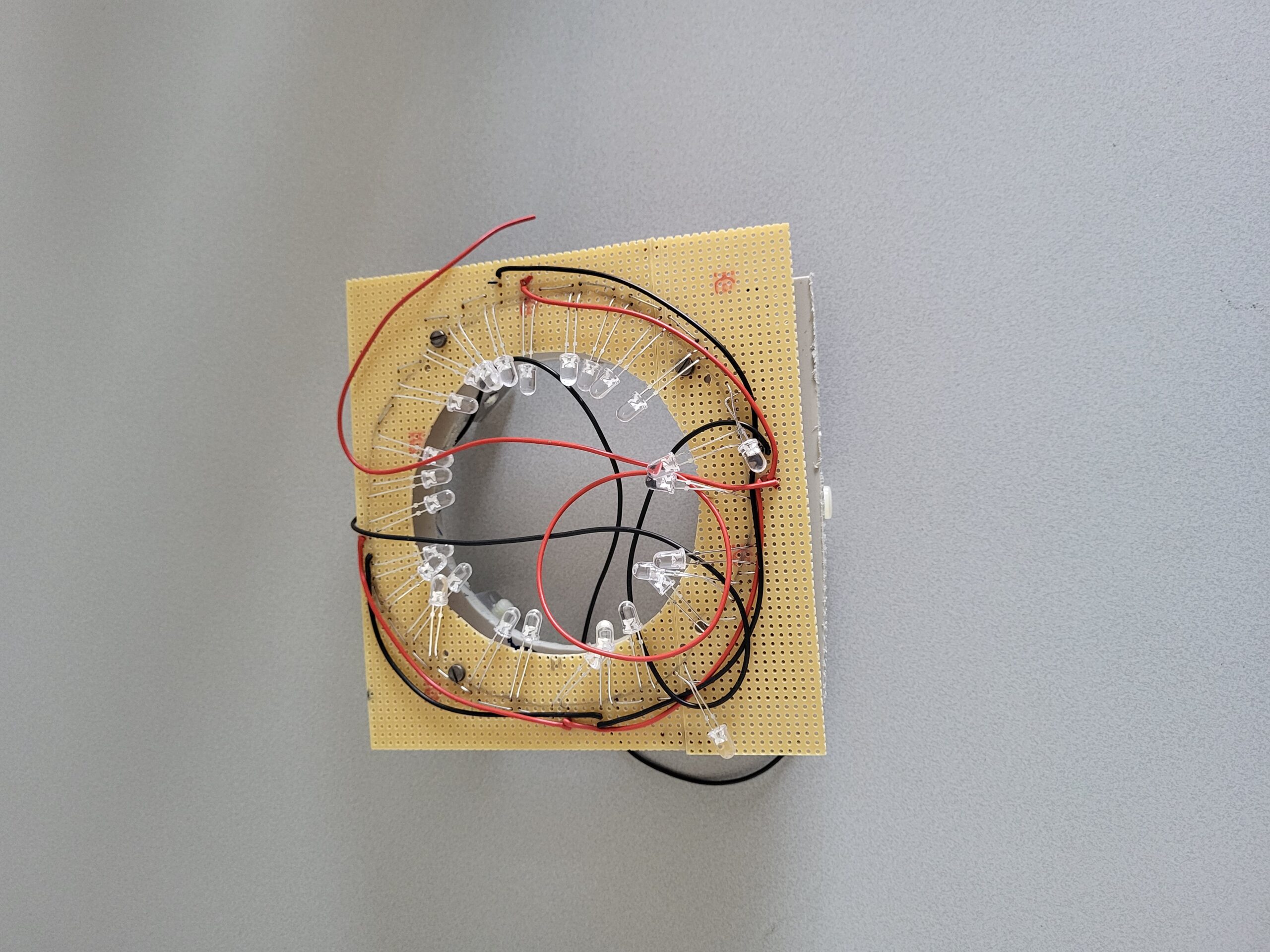
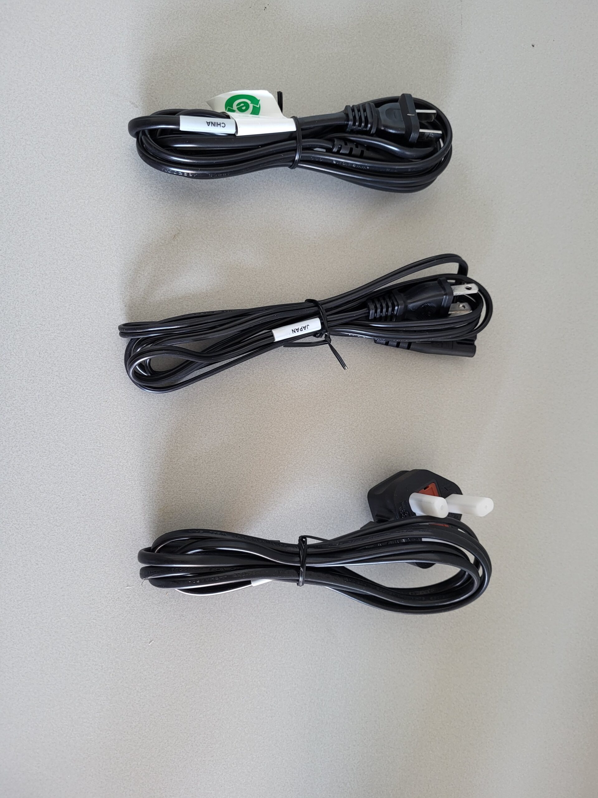
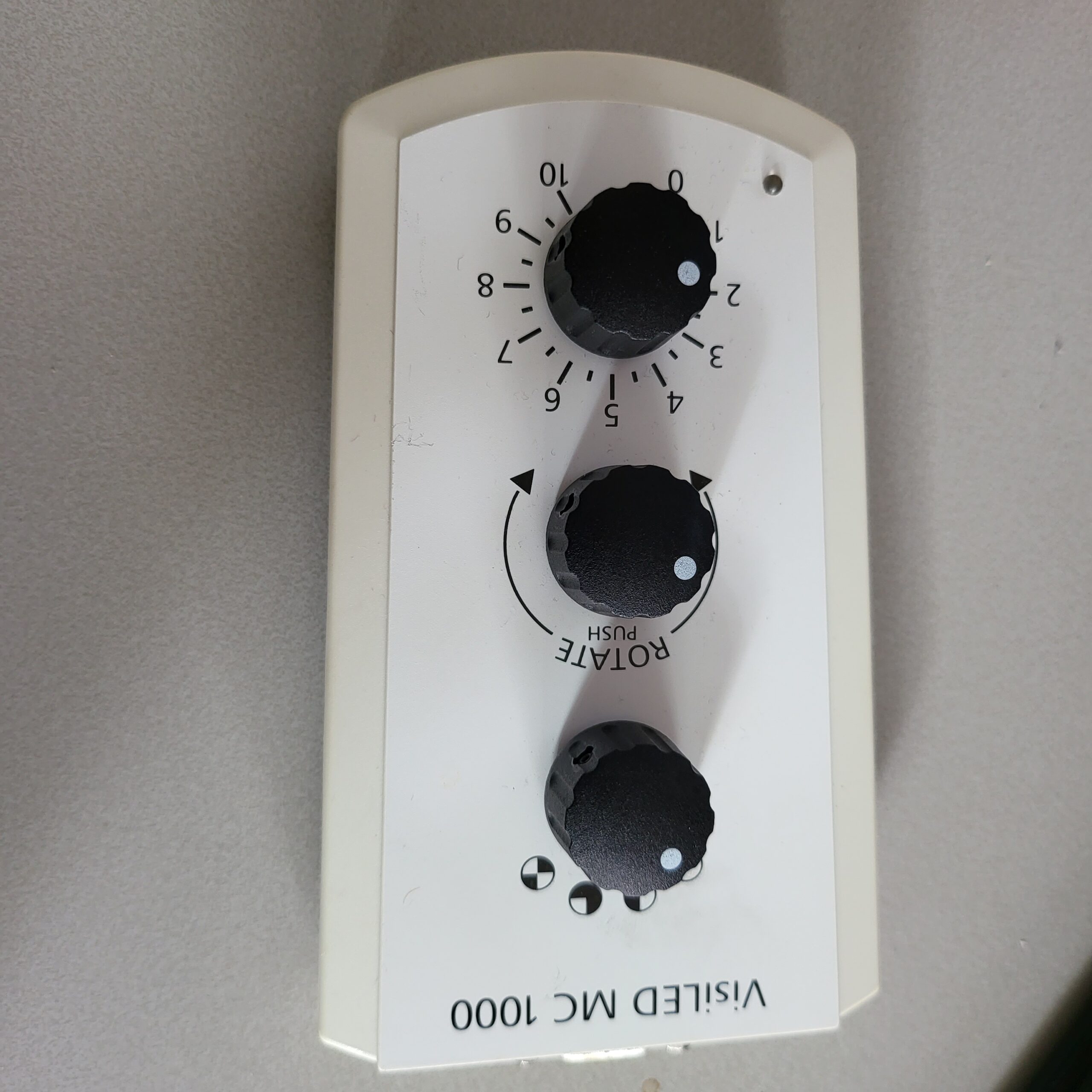
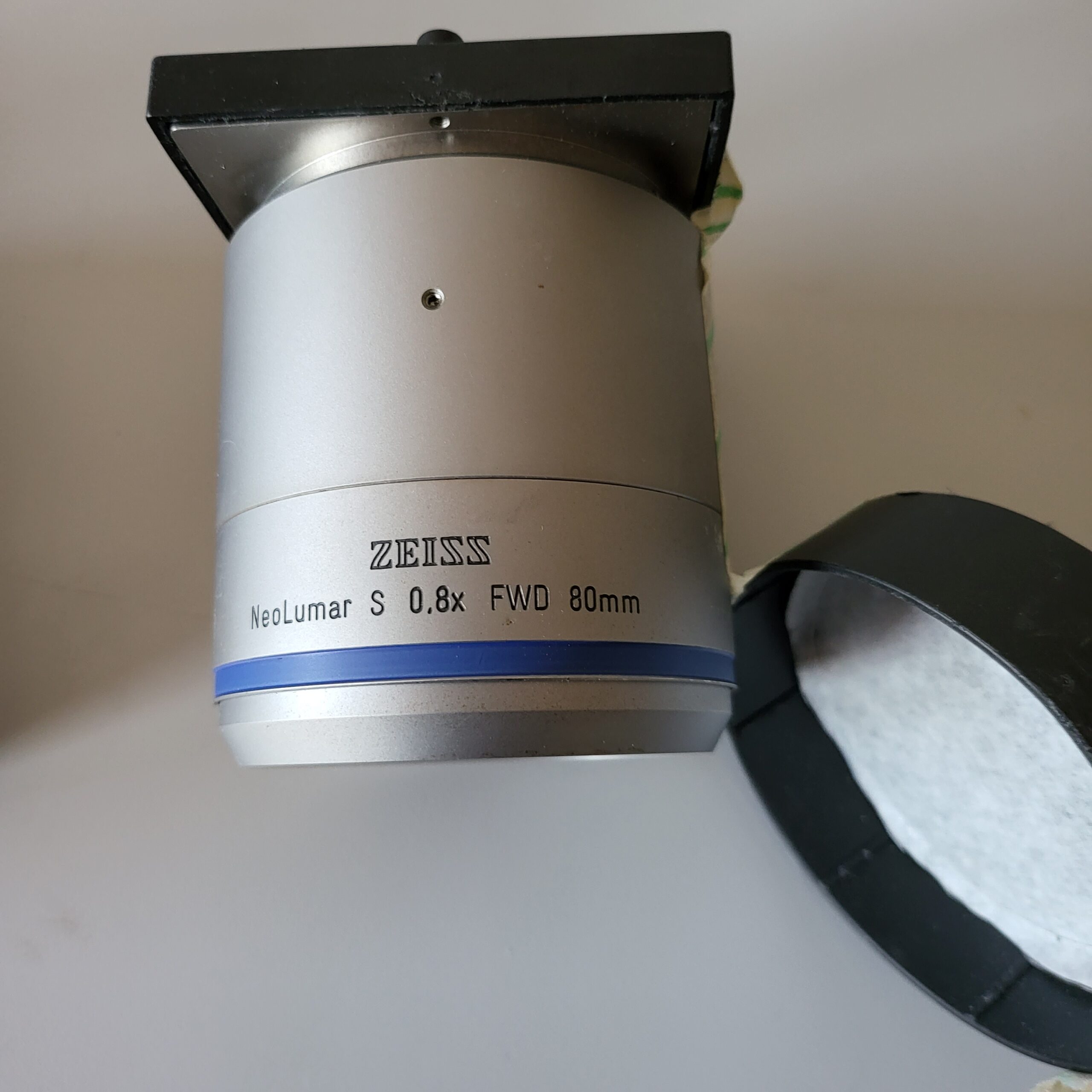
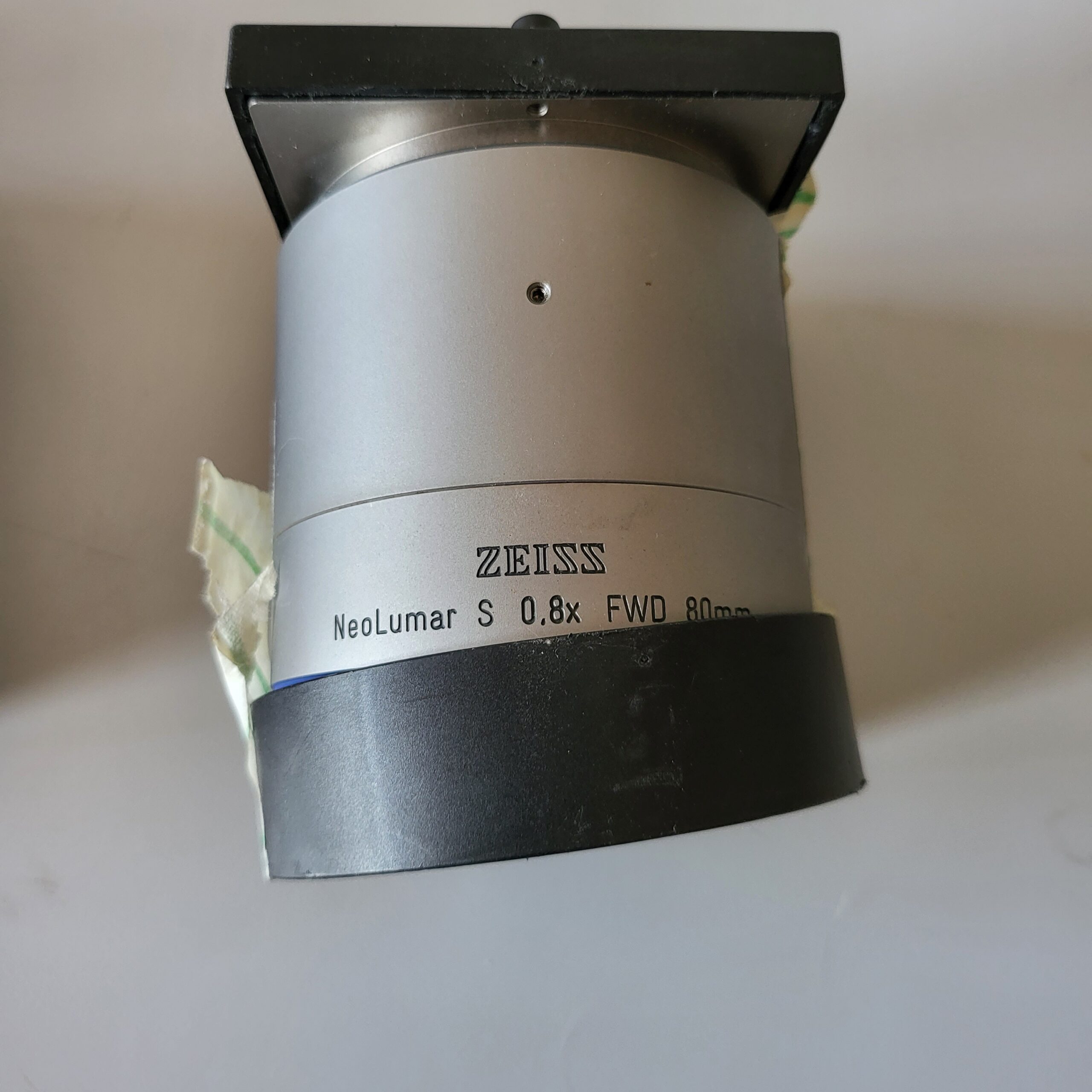
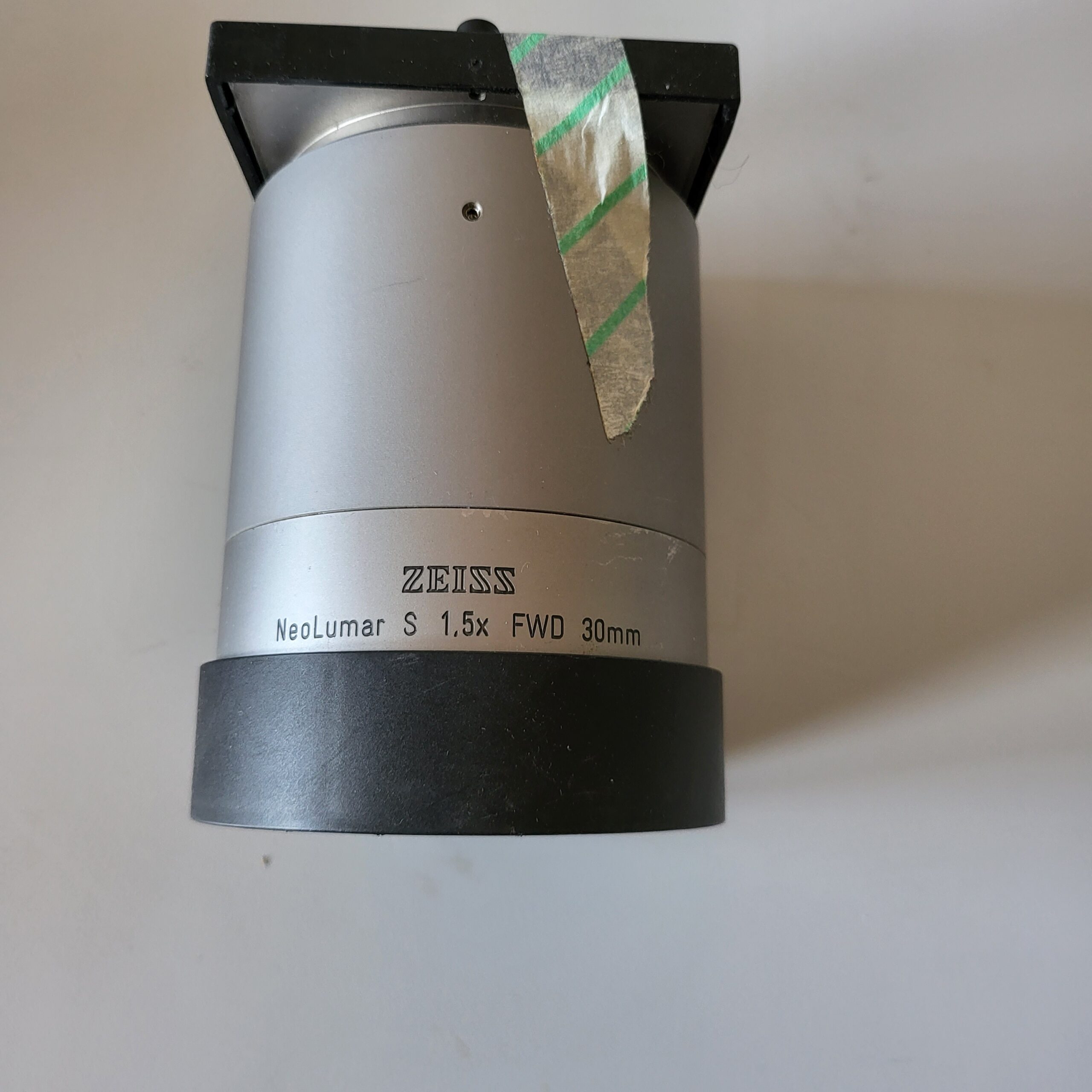
This Used Zeiss Stereo Lumar V12 with HBO 100 Illuminating system was tested to certain degree to make sure all mechanical parts are working. The system is missing the Neolumar S objective with dovetail guide and the KL 2500 LCD cold light source. Everything else is included according to the pictures. Other comparable system are sold for around $20.000. Non working systems are sold for $8000 so we offer a cheaper alternative for someone willing to buy some parts separate.
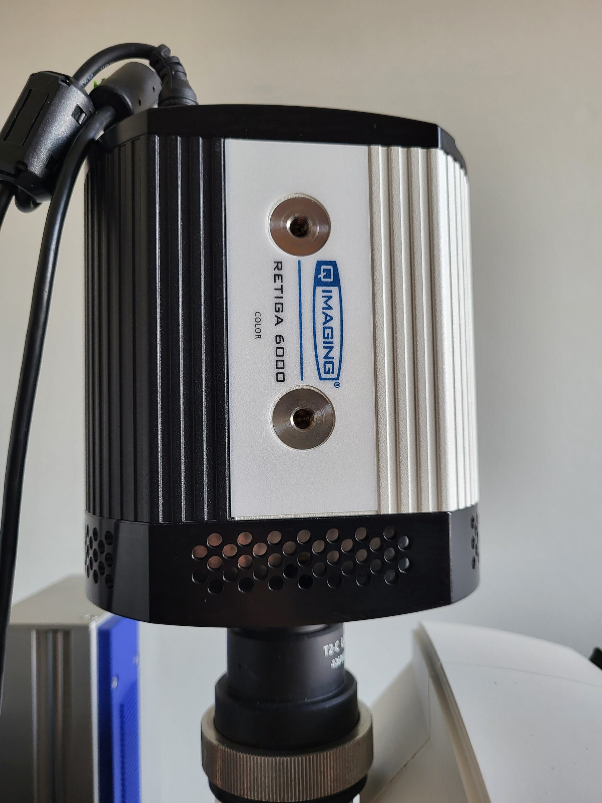
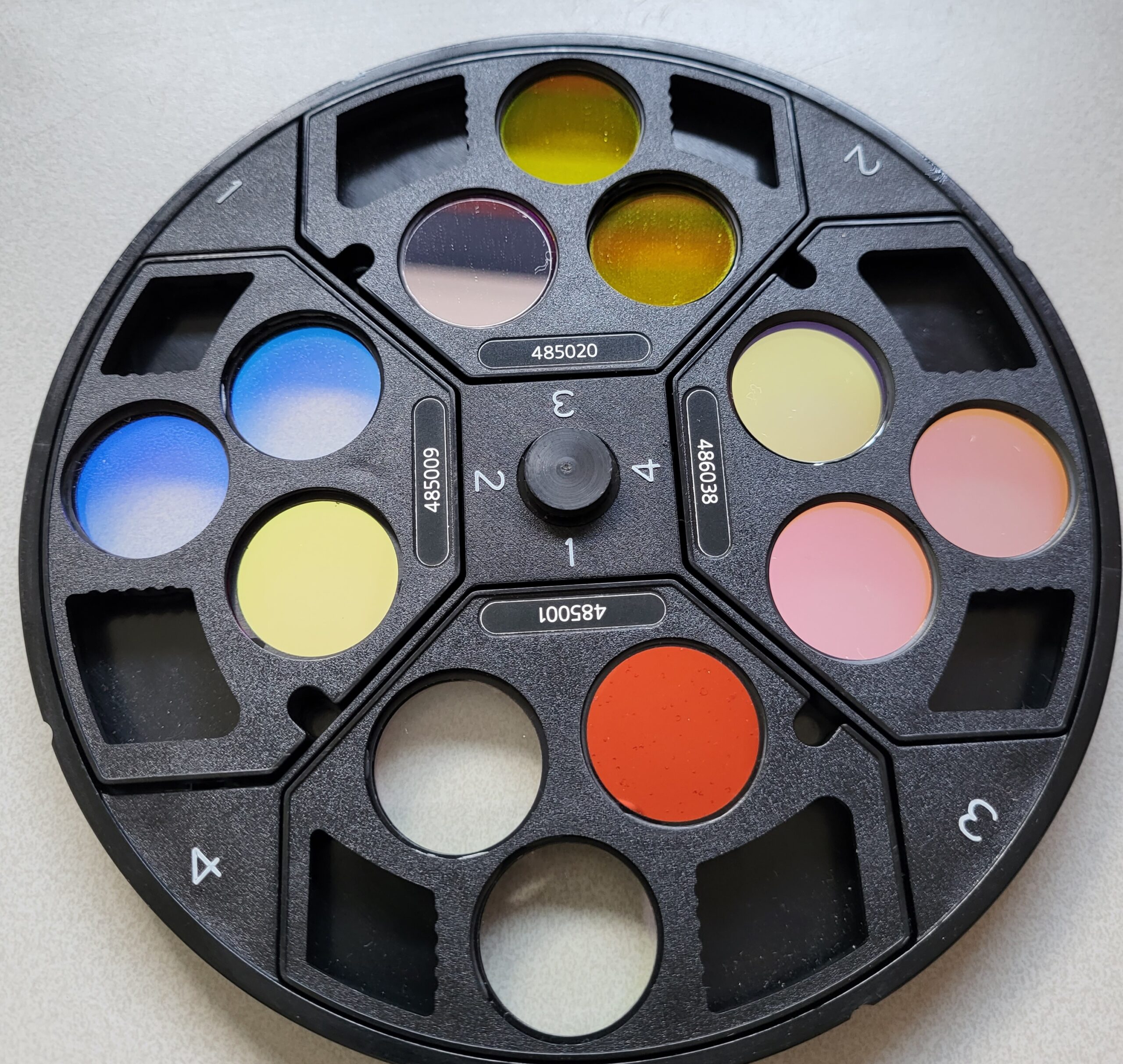
The Zeiss Lumar V12 is a fluorescence stereo microscope suitable for high resolution 3D analysis of large specimens in fluorescent and transmitted light. The system is equipped with fluorescence filters for GFP, Cy3 and Cy5.5 and a sensitive cooled CCD camera that allows you to capture weak fluorescent signals and minimize photobleaching in light sensitive samples. With the Zeiss Lumar V12 you can perform multi-channel, Z-stack and time-lapse image acquisition of your samples, using two different objectives with different working distances and magnification values. If you need higher magnifications, check out the Leica DM5000B, the Zeiss Axiovert 200M or the Zeiss Cell Observer widefield microscopes.
The Zeiss Lumar V12 is a fluorescence stereo microscope suitable for high resolution 3D analysis of large specimens in fluorescent and transmitted light. The system is equipped with fluorescence filters for GFP, Cy3 and Cy5.5 and a sensitive cooled CCD camera (Hamamatsu ORCA-R2) that allows you to capture weak fluorescent signals and minimize photobleaching in light sensitive samples. With the Zeiss Lumar V12 you can perform multi-channel, Z-stack and time-lapse image acquisition of your samples, using two different objectives with different working distances and magnification values. If you need higher magnifications, check out the Leica DM5000B, the Zeiss Axiovert 200M or the Zeiss Cell Observer widefield microscopes.
Reproducible Results Over the Motorized 12:1 Zoom Range
The motorized components of your SteREO Discovery.V12 are fully integrated into your microscope software ZEN and AxioVision: measure and document your samples reproducibly. Observe your Drosophila or zebrafish embryos, stents or circuit boards with an enhanced threedimensional impression over the whole 12:1 zoom range.
Capture images that are crisp and rich in contrast, have excellent color fidelity and simply contain more information. SteREO Discovery.V12 enhances resolution and contrast in biology and quality assurance.
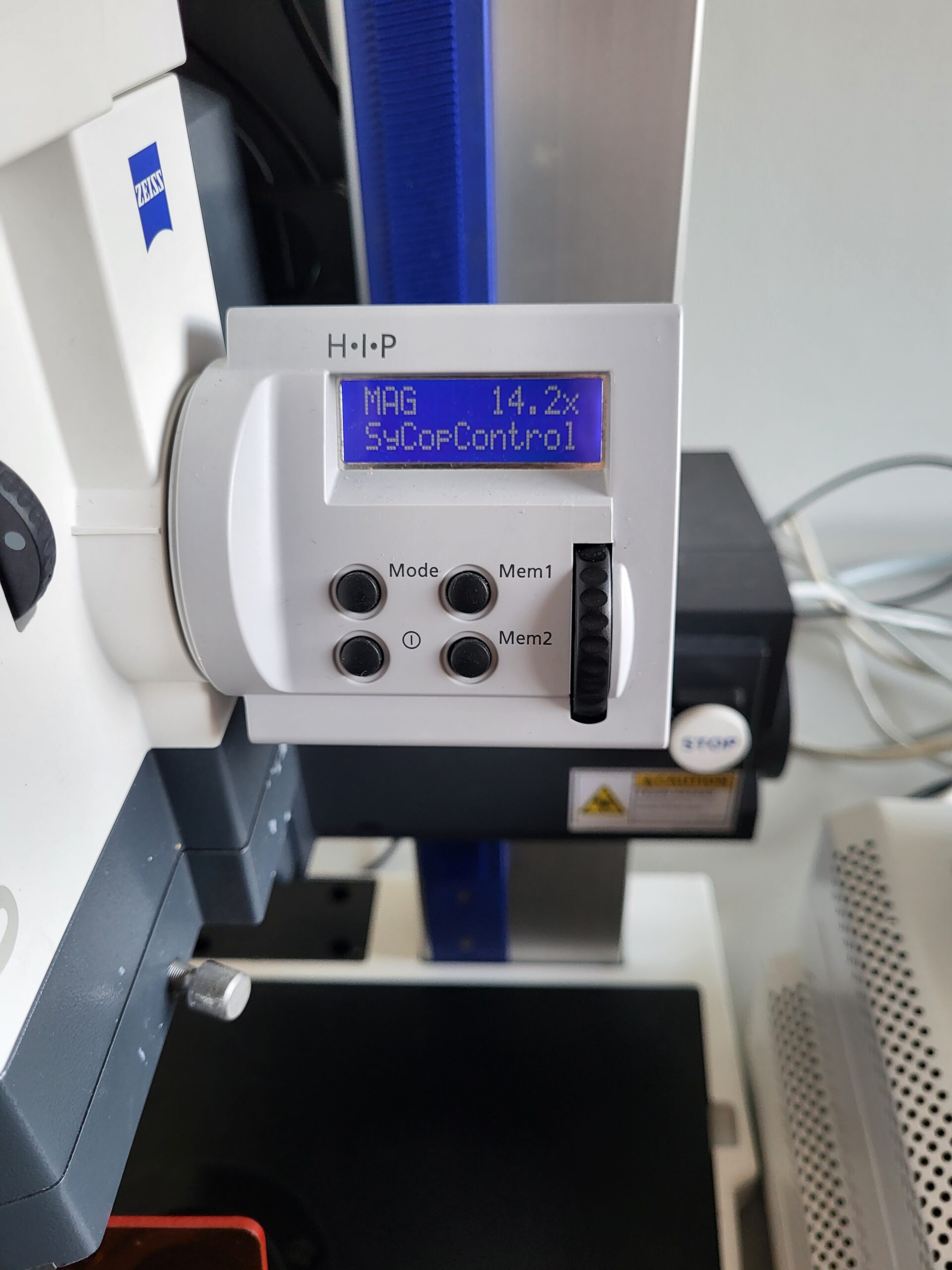
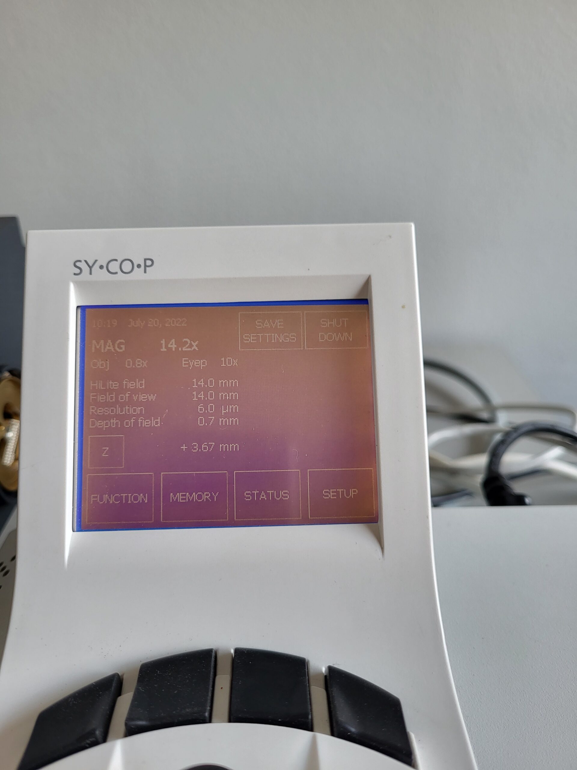
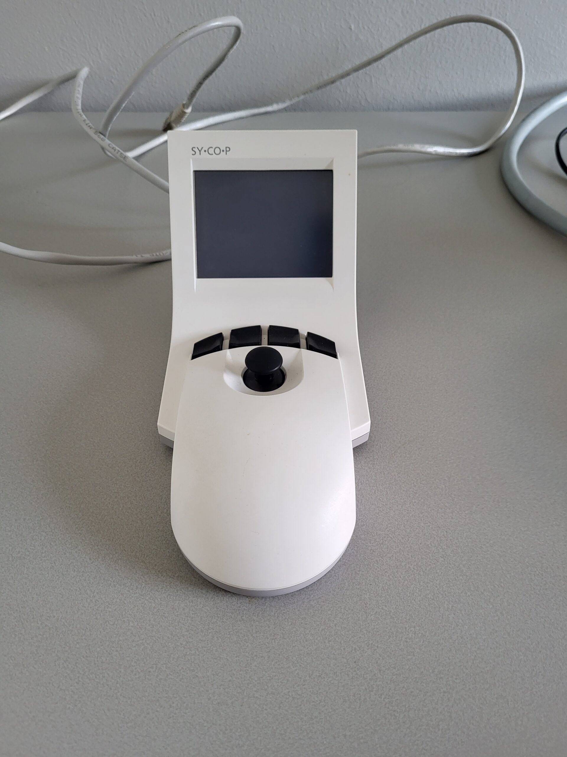
The external touch panel SYCOP controls all essential functions of SteREO Discovery.V12: magnification, focus, contrast, brightness and more.
Highlights
The optical concept
Carl Zeiss, optical pioneer and inventor of stereomicroscopy, has invested its entire know-how and expertise in the development of a new optical design for SteREO.Discovery.V12 – with remarkable
success. The patented optics are setting new standards in modular common main objective stereomicroscopy. Thanks to higher resolution and significantly increased contrast, they provide 20%
more image information – in addition to sensational 3D images.
Flexibility: Intelligent interfaces
The modular design of SteREO Discovery.V12, typical of common main objective stereomicroscopes, offers greater flexibility in the selection of components. In addition, intelligent interfaces
enable the use of diverse options from the comprehensive illumination and accessory program from Carl Zeiss
Lumar V12 Fluorescence
Developed for conventional light fluorescence microscopy, SteREO Lumar.V12 offers first-class optical illumination systems These systems feature
even illumination of specimens at all magnifications, and an independent zoom for optimal adaptation of fluorescence excitation light to the specimen field selected. In addition, continuous automation makes the handling of filters particularly easy.
For optimal fluorescence excitation, the integrated light zoom with lamp mount and 4x filter mount (HiLite) is automatically coupled to the observation
zoom. A remarkable option: the light zoom can be detached from the observation zoom. The advantage of the free light zoom: it is possible to
individually increase the brightness of selected specimen structures at low resolutions – thereby mobilizing the last reserves of light. And at high
resolutions the light intensity can be reduced, thus ensuring deblurred specimens.
Includes:

